
Topics
What Is an Enlarged Heart? The Parts of the Heart Diagnosing an Enlarged Heart: The Critical Role of Each Examination Causes of an Enlarged Heart Treatment and Management Strategies Prevention of an Enlarged Heart ConclusionReceiving information from a doctor or reading a medical report that mentions an "enlarged heart on an X-ray" can certainly cause anxiety and raise many questions. What does this mean for your health? How serious is the condition? And what are the next steps to take?
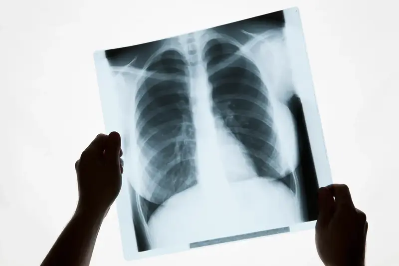
It's true that a chest X-ray (also called a chest radiograph) is often the first gateway to detecting a potential enlargement of the heart, known medically as cardiomegaly. However, it is crucial to understand that this X-ray result is not a final diagnosis but rather a very valuable initial clue. It serves as a signal for the doctor to conduct a more thorough investigation.
To truly understand the heart's condition in detail, including the precise size of each heart chamber, the thickness of the muscle, the function of the valves, and its pumping strength, more advanced and accurate examinations are required. This article will provide a comprehensive overview of everything you need to know, from what an enlarged heart on an X-ray finding means and why it happens, to the proper diagnostic and treatment pathway according to medical standards.
What Is an Enlarged Heart?
An enlarged heart, or cardiomegaly, is a condition where the heart's size is larger than normal. This occurs when the heart muscle enlarges or its walls thicken. This condition is typically caused by various factors that affect the heart's function and workload. The enlargement can be temporary or permanent, depending on the underlying cause. In some cases, an enlarged heart can still function normally, but in more severe conditions, it can reduce the efficiency of blood pumping and increase the risk of heart failure.
There are two main types of cardiomegaly based on their mechanism of enlargement. First is cardiac dilation, which occurs when the heart's chambers stretch and enlarge due to excess pressure, often seen in cases of heart failure or dilated cardiomyopathy. Then, there is cardiac hypertrophy, which occurs when the heart muscle walls thicken due to high blood pressure or other diseases that force the heart to work harder. While an enlarged heart can be suggested by an X-ray, confirming its size and assessing its pumping function requires an echocardiogram.
Meanwhile, the symptoms of an enlarged heart will appear according to its severity and can significantly affect a patient's activities. One of the main symptoms is shortness of breath, especially during activity or when lying down, due to the heart's inability to pump blood effectively. Additionally, patients may experience an irregular heartbeat (arrhythmia), which can cause palpitations or dizziness. Chest pain is also common, especially if blood flow to the heart is compromised. Fluid retention due to impaired blood circulation can lead to swelling in the legs and ankles. Another common symptom is fatigue or weakness, as the body does not receive an adequate supply of oxygen and nutrients from suboptimal blood circulation.
The Parts of the Heart
Before discussing an enlarged heart on an X-ray further, it's important to know the parts of the heart. The heart consists of several main parts, each with a specific function in the process of blood circulation. Every part plays a vital role in directing blood flow. Here are the parts of the heart you should know:
- Right Atrium: Receives carbon dioxide-rich blood from the entire body through the superior and inferior vena cava.
- Right Ventricle: Pumps blood to the lungs through the pulmonary artery for oxygen exchange.
- Left Atrium: Receives oxygen-rich blood from the lungs through the pulmonary veins.
- Left Ventricle: Pumps oxygen-rich blood to the entire body through the aorta.
- Heart Valves: Consist of several parts such as the tricuspid, pulmonary, mitral, and aortic valves.
- Aorta: The largest artery that carries oxygen-rich blood to the entire body.
- Coronary Arteries and Veins: Provide the blood supply to the heart muscle to maintain its function.
- Sinoatrial (SA) Node and Atrioventricular (AV) Node: Control the heartbeat and ensure that heart contractions occur regularly.
Diagnosing an Enlarged Heart: The Critical Role of Each Examination
Diagnosing cardiomegaly is a systematic investigation process, starting from initial findings to a definitive confirmation.
- Initial Clues from an Enlarged Heart X-Ray Result
A chest X-ray is a quick, non-invasive, and widely available screening test. During the X-ray, the doctor analyzes the heart's silhouette or shadow. One simple metric used is the Cardiothoracic Ratio (CTR). This is a comparison between the horizontal width of the heart and the internal width of the chest cavity (thorax). If this ratio is greater than 0.5 (or 50%), it indicates the presence of an enlarged heart on the X-ray.
Although very useful as an initial marker, it is important to be aware of the limitations of an X-ray:
- Two-Dimensional Image: An X-ray only provides a two-dimensional shadow. It cannot distinguish between an enlargement of the heart muscle (hypertrophy) and the presence of fluid in the sac surrounding the heart (pericardial effusion), both of which can make the heart's silhouette appear larger.
- Does Not Assess Function: An X-ray provides absolutely no information about the heart's pumping function, the condition of the valves, or blood flow.
- Technical Variability: The results can vary slightly depending on the patient's position and the degree of inspiration (how deeply they breathe in) when the image is taken.
So, if you receive a result showing an enlarged heart on an X-ray, do not panic. Consider it a yellow flag, an important signal to proceed to the next stage of examination.
- Confirming the Diagnosis with an Echocardiogram (Heart Ultrasound)
This is the gold standard and most crucial examination in the evaluation of an enlarged heart. An echocardiogram uses sound waves (ultrasound) to create real-time, moving images of the heart. The procedure is painless; a doctor will apply a gel to your chest and move a device called a transducer over it. The advantages of an echocardiogram are truly remarkable:
- Accurate Measurements: It can precisely measure the thickness of the heart walls and the dimensions of each heart chamber in millimeters.
- Pumping Function Analysis: It provides a vital metric called the Ejection Fraction (EF). EF is the percentage of blood pumped out of the left ventricle with each heartbeat. A normal EF value is around 55-70%. A value below that indicates a weakening of the pumping function.
- Valve Evaluation: It allows for direct visualization of how the four heart valves open and close, detecting any leakage (regurgitation) or narrowing (stenosis).
- Blood Flow Visualization: With Doppler technology, the doctor can see the direction and speed of blood flow within the heart.
The results from the echocardiogram are what will provide a definitive diagnosis and help the doctor understand the type and severity of the heart enlargement.
- Other Supporting Tests to Find the Cause
Once cardiomegaly is confirmed, the next step is to find out the cause.
- Electrocardiogram (ECG): Records the heart's electrical activity to detect arrhythmias, signs of heart muscle thickening, or evidence of a past heart attack.
- Blood Tests: Can detect markers like BNP (B-type natriuretic peptide), which increases in heart failure, and check for other potential causes like anemia, thyroid disorders, or kidney problems.
- Stress Test: The patient is asked to walk on a treadmill while their ECG and blood pressure are monitored to look for signs of coronary artery disease (blocked blood vessels).
- Heart CT Scan or MRI: Provides highly detailed images of the heart's structure, coronary blood vessels, and can detect scar tissue on the heart muscle.
- Cardiac Catheterization: An invasive procedure where a thin tube is inserted through a blood vessel in the arm or groin up to the heart to measure pressures inside the heart and directly visualize blockages in the coronary arteries.
Causes of an Enlarged Heart
After identifying an enlarged heart on an X-ray, it is essential to know the potential causes. An enlarged heart can be caused by various medical conditions that increase the heart's workload. For example, coronary artery disease occurs due to blockage of the coronary arteries, reducing blood flow to the heart and causing muscle damage and enlargement. Additionally, cardiomyopathy is a disorder of the heart muscle that causes weakness, forcing the heart to work harder to pump blood. Heart valve disorders can also be a contributing factor, where improperly functioning valves allow blood to flow backward, increasing pressure and the heart's workload.
Besides factors directly related to the heart, several other conditions can also trigger heart enlargement. Arrhythmia, for example, disrupts the regularity of the heartbeat, which can affect pumping efficiency. Chronic lung diseases such as COPD and pulmonary hypertension also contribute by increasing pressure on the heart, leading to enlargement. Chronic anemia, characterized by a lack of red blood cells, can also cause long-term heart enlargement by forcing the heart to work harder to supply oxygen to the entire body.
Treatment and Management Strategies
The goal of treatment is to address the underlying cause, improve symptoms, and prevent the condition from worsening.
- Fundamental Lifestyle Changes: This is the foundation of all treatment. It includes a low-salt diet (to reduce fluid retention and blood pressure), quitting smoking, limiting or stopping alcohol consumption, managing stress, and engaging in light to moderate physical activity as advised by a doctor.
- Medication Therapy:
- ACE Inhibitors or ARBs: Cornerstone medications to relax blood vessels, lower blood pressure, and reduce the heart's workload.
- Beta-blockers: Slow the heart rate, lower blood pressure, and in the long term, can help improve the heart's shape and function.
- Diuretics: Help the body excrete excess salt and water through urine, thereby reducing swelling and shortness of breath.
- Anticoagulants (Blood Thinners): May be necessary if there is a risk of blood clot formation in the enlarged heart chambers, especially in patients with atrial fibrillation.
- Medical and Surgical Procedures:
- Pacemaker or ICD (Implantable Cardioverter-Defibrillator) Implantation: To manage dangerous heart rhythms.
- Coronary Intervention: Such as stent placement or bypass surgery to address blockages in the coronary arteries.
- Valve Surgery: To repair or replace damaged heart valves.
- Heart Transplant: As a last resort for end-stage heart failure that does not respond to other treatments.
Prevention of an Enlarged Heart
The results of an enlarged heart X-ray can also help determine preventive measures to stop the condition from worsening. Maintaining heart health can be achieved through various preventive steps. Regularly checking blood pressure and cholesterol levels can help detect the risk of heart disease early.
Furthermore, avoiding excessive stress also plays a role in maintaining heart health, as prolonged stress can trigger an increase in blood pressure and irregular heartbeats. Maintaining an ideal body weight is another important factor, as excess weight can burden the heart and increase the risk of cardiovascular disease. Additionally, controlling blood sugar levels is necessary to prevent diabetes, which can trigger various heart complications if not well-managed.
Conclusion
Seeing an enlarged heart on your medical report is an important starting point, a signal that your heart needs more attention. It is not a final verdict, but rather an invitation to undergo a more comprehensive evaluation.
The correct diagnostic process involves a series of tests, from an X-ray to an echocardiogram, to provide a detailed picture of the heart's structure and function. By knowing the exact cause, your doctor can design the most effective treatment plan for you. Modern medicine, combined with a healthy lifestyle, can significantly improve symptoms, enhance quality of life, and slow the progression of the disease.
